

Last updated on 24 August 2026
Rift Valley fever (RVF) is an arboviral infection.¹ It was first isolated in 1931 during an epizootic outbreak among sheep in Kenya’s Rift Valley.² The first major human epidemic occurred in Egypt in 1977, when between 18,000 and 200,000 cases were reported, resulting in approximately 600 deaths.¹
Endemic in sub-Saharan Africa and the Middle East, RVF outbreaks typically occur following periods of exceptionally heavy rainfall that promote mosquito proliferation.¹˒² In France, Mayotte recorded ten confirmed human cases of RVF in 2007, mainly associated with contact with ruminants.³ In 2018–2019, the archipelago reported 142 confirmed cases, 73% of which were linked to animal exposure.⁴ The outbreak was successfully controlled through veterinary investigations, vector control measures and targeted prevention efforts.
The RVF virus is an arbovirus, meaning a virus transmitted through the bite of blood-feeding arthropod vectors. This single-stranded RNA virus belongs to the genus Phlebovirus, within the family Phenuiviridae and the order Bunyavirales.¹˒⁵˒⁶
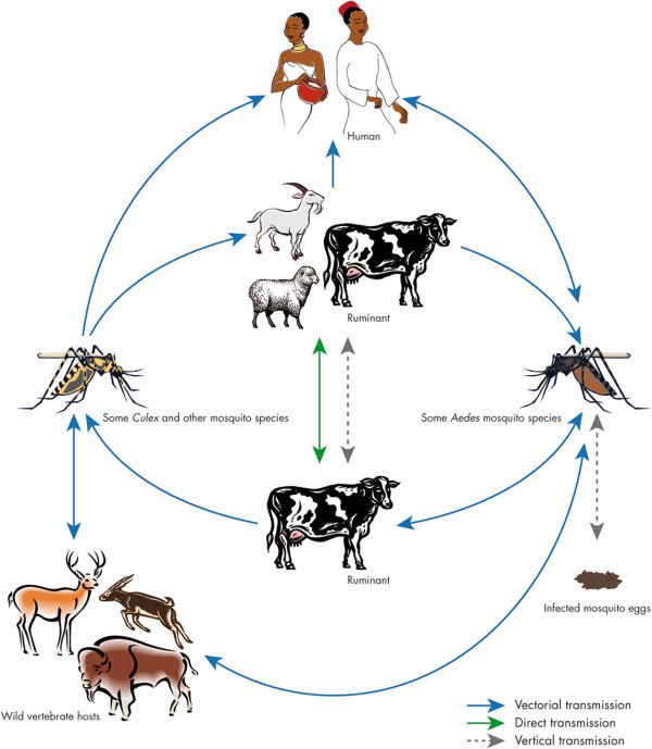
RVF primarily affects ruminants (sheep, cattle, goats and camels).⁷ Vector-borne transmission through the bite of infected mosquitoes (mainly Aedes and Culex species, including A. albopictus and C. pipiens) plays a major role in virus transmission among animals but appears to play a lesser role in transmission to humans.⁷
The virus persists in primary foci through an Aedes–animal cycle involving vertical transmission within mosquito populations. Major outbreaks spread to secondary foci through livestock movements, wildlife reservoirs or vector dispersal, and are subsequently amplified among ruminants by Culex mosquitoes in irrigated areas. Epizootics consistently precede human epidemics.⁷
In most cases, infection results from direct or indirect contact with blood or organs from infected animals during slaughtering or butchering, animal births and veterinary procedures, or when handling carcasses or aborted foetuses.⁷ Transmission may also occur through the consumption of raw or unpasteurised milk from infected animals.⁷
To date, no human-to-human transmission has been documented.⁷
RVF can be clinically difficult to distinguish from other infectious diseases such as malaria, dengue, yellow fever or other viral haemorrhagic fevers.⁷ Symptoms including fever, headache, muscle pain and, in some cases, haemorrhagic or neurological manifestations may resemble those of other systemic infections.
RVF diagnosis can be confirmed using several laboratory methods:⁶˒⁷
In animals, RVF causes widespread abortions and high mortality rates.²˒⁷
In humans, most infections are asymptomatic. Among symptomatic cases, the most frequently reported clinical manifestations are fever (81%), renal failure (41%), nausea (38%) and encephalitis (21%). Death occurs in 21% of cases; most patients who died had been hospitalised.⁸
The incubation period ranges from two to six days.⁷
There is currently no specific treatment available for RVF virus infection. In most human cases, where the disease is relatively mild and self-limiting, no specific treatment is required. Management is based on supportive and symptomatic care aimed at relieving fever and pain and preventing hepatic or ocular complications.⁷˒⁹
For more severe cases, treatment relies primarily on early intensive supportive care, including rehydration and symptom management.⁷
Research is underway to identify antiviral compounds active against RVF virus, including molecules already approved for other viral diseases. Certain antivirals, such as ribavirin, favipiravir and viral replication inhibitors, have shown partial efficacy in vitro or in animal models, although none has yet demonstrated sufficient clinical efficacy in humans.¹⁰
The results obtained with two monoclonal antibodies capable of providing post-exposure protection in mouse models offer promising prospects for future targeted immunotherapy in humans.¹¹
Prevention primarily relies on non-pharmaceutical measures aimed at limiting virus transmission. This includes reducing the risk of animal-to-human transmission through the use of gloves and personal protective equipment when handling animals or biological animal products.
Individual and community protection against mosquito bites is also essential and includes insecticide-treated bed nets, protective clothing, insect repellents and improvements to housing conditions. Vector control also plays a key role, particularly through the management of standing water, targeted insecticide use and entomological surveillance in at-risk areas.⁷˒⁹
To date, only one vaccine has been used in humans. This is the inactivated vaccine TSI-GSD-200, developed by the United States Army. However, it is not commercially available and remains experimental, being reserved for the protection of veterinarians and laboratory personnel at high risk of exposure.²˒¹²
Several vaccine candidates are currently under development, including the inactivated vaccine TSI-GSD-200 and the live-attenuated vaccine MP-12, both of which have demonstrated good immunogenicity. Both are currently being evaluated in Phase II clinical trials.¹³
The University of Oxford, through a project supported by the Coalition for Epidemic Preparedness Innovations (CEPI), has developed the ChAdOx1 RVF vaccine. This candidate uses an adenoviral vector platform similar to that employed in the development of certain Covid-19 vaccines. Preclinical and Phase I studies have demonstrated a favourable safety profile, strong immunogenicity and promising thermostability. The vaccine is currently being evaluated in Kenya in a Phase II clinical trial conducted by the KEMRI-Wellcome Trust Research Programme, making it one of the most advanced candidates for human use in endemic regions.
The vaccination approach for RVF has evolved considerably. It now incorporates strategies allowing vaccinated individuals to be distinguished from naturally infected individuals through serological testing (DIVA – Differentiating Infected from Vaccinated Animals). Several next-generation vaccine candidates currently under development, including RVFV-4s, DDVax, ChAdOx1-RVF and MP12, are vector-based vaccines intended for both human and veterinary use. Among these, RVFV-4s, a four-segmented virus, and DDVax, a virus containing two genetic deletions, have been specifically designed to maximise safety. Because they are incapable of further propagation, they reduce vaccination-related risks while enabling accurate serological monitoring.
Promising results suggest that these vaccines could provide effective protection in humans, reduce transmission risks and facilitate outbreak management, which would be crucial for improving prevention of this zoonotic disease in regions at risk.¹³
Through its involvement in the WHO CORC network (including coordination of the Filovirus CORC), ANRS MIE contributes to discussions on international research priorities for Rift Valley fever.
These discussions facilitate coordination with other research stakeholders, monitoring of ongoing work, identification of emerging advances and recognition of areas where additional efforts are required, thereby supporting the development of complementary projects and strengthening the coherence of the global scientific response.
Arbo-France, coordinated under the auspices of ANRS Emerging infectious diseases, is a French network dedicated to the study of arboviral diseases. Its objective is to facilitate preparedness for and response to human and animal arbovirus outbreaks in mainland France and overseas territories.
ANRS Emerging infectious diseases activated a Level 1 outbreak response unit on 11 December 2025 in response to an outbreak in Senegal and Mauritania. The cell has since been stood down.
Regular scientific monitoring updates were shared with members of the agency’s network, and a research meeting was held on 7 November 2025, bringing together experts from Senegal and Mauritania. The objectives of this meeting were to review the epidemiological situation in both countries, identify major challenges to the response, define priority research areas and foster collaborative initiatives. Several research priorities emerged from these discussions: