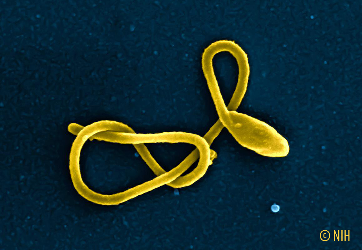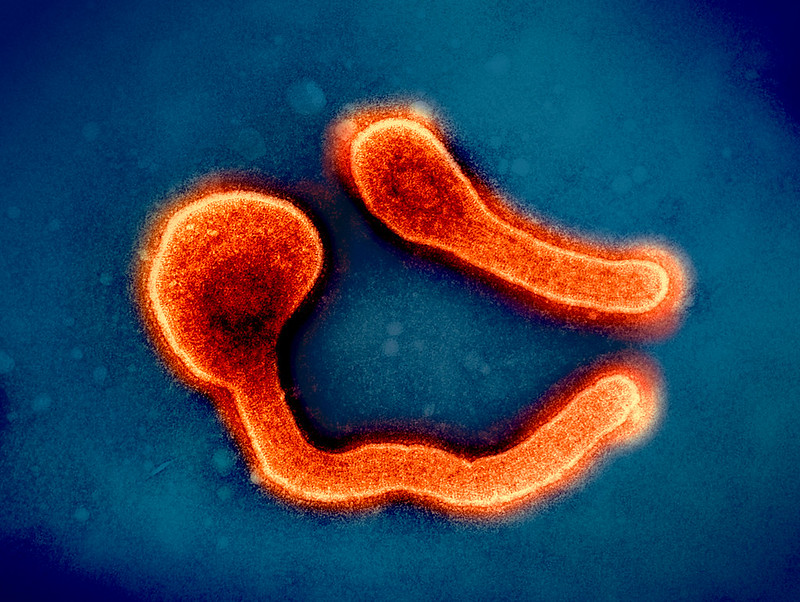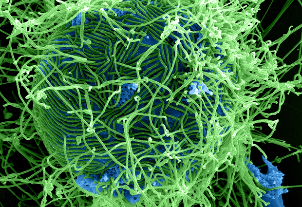
Viral haemorrhagic fevers (VHFs) encompass various diseases (Ebola, Lassa fever, Marburg fever, Crimean–Congo fever, etc.).
Last updated on 24 August 2026
Yellow fever is a centuries-old example of a viral haemorrhagic fever (VHF).⁶ Ancient China, Manchuria in 1930, countries in north-western Europe in 1934 and Korea in the 1950s experienced, under various names, an acute epidemic viral haemorrhagic fever associated with acute kidney injury.⁶ In 1962, Gajdusek grouped these clearly related infections together under the term haemorrhagic fever with renal syndrome (HFRS), caused by viruses belonging to the same genus, Hantavirus, within the Bunyaviridae family.⁶
It subsequently became apparent that VHFs form a heterogeneous group of viral diseases involving different virus families.⁶˒⁷ The emergence and re-emergence of viral haemorrhagic fevers are a growing global concern.⁸
Over the past 20 years, the Eastern Mediterranean Region has experienced major outbreaks and sporadic cases of yellow fever, Rift Valley fever, severe dengue and Crimean–Congo haemorrhagic fever in more than 12 countries.⁸ The Ebola epidemic that occurred in West Africa in 2015 resulted in more than 29,000 cases and approximately 11,000 deaths.⁹
The biology of the viruses responsible for VHFs, their epidemiology and their case fatality rates vary considerably: 1% for Lassa fever,¹ 50% for Ebola²˒³ and Marburg haemorrhagic fever,⁴ and 40% for Crimean–Congo fever.⁵
The unprecedented mobilisation that followed the Ebola epidemic highlighted the need to implement structuring research initiatives to improve preparedness for viral haemorrhagic fever epidemics, which pose public health risks because of their epidemic potential and/or the absence or inadequacy of countermeasures.
Different virus families are involved in the development of VHFs:⁶˒⁷
Find out more about:
Filoviruses were discovered in 1967 following the emergence of a severe haemorrhagic syndrome in Marburg, Germany, and Belgrade, Serbia.¹⁰ The virus identified at that time was named Marburg virus.¹⁰ In 1976, two VHF outbreaks occurred, one in South Sudan and the other in the Democratic Republic of the Congo.
These respectively represented the first descriptions of Sudan virus and Ebola virus.¹⁰ Subsequently, with the exception of a few sporadic cases, filoviruses did not reappear in humans for 18 years before re-emerging in the form of epidemic outbreaks.¹⁰
In 2013, Ebola virus disease emerged in Forested Guinea, spread to Liberia and Sierra Leone and eventually developed into an outbreak in West Africa, causing the most devastating filovirus epidemic to date, with more than 29,000 cases and approximately 11,000 deaths between December 2013 and June 2016.⁹˒¹⁰
Since then, various diseases caused by filoviruses have been reported, including Marburg virus disease and Ebola virus disease caused by either Ebola virus or Sudan virus.¹⁰
To date, outbreaks caused by ebolaviruses have been confined to African countries, but this could change, as imported cases have been reported in Europe and the United States.⁷˒¹¹
Filoviruses include two genera that are pathogenic to humans: Ebolavirus and Marburgvirus.¹⁰ The genus Ebolavirus comprises six species. Three of these are highly pathogenic to humans: Zaire ebolavirus (Ebola virus), Bundibugyo ebolavirus (Bundibugyo virus) and Sudan ebolavirus (Sudan virus).¹⁰
Virions are enveloped and vary in morphology. Their genome consists of a negative-sense, single-stranded RNA molecule encoding seven proteins.¹⁰ In ebolaviruses, the viral gene encodes a protein secreted by infected cells, sGP, or secreted glycoprotein. sGP is transformed into a structural glycoprotein that plays an important role in virulence, although other pathogenicity factors also exist.¹⁰
VHFs have multiple modes of transmission.⁸ Transmission may occur through contact with carrier animals, such as carcasses or fluids from infected animals in the case of Lassa, Marburg or Ebola fevers; through insect bites, including mosquitoes and ticks in the case of Crimean–Congo and Rift Valley fevers; and through human-to-human transmission involving blood, secretions and other bodily fluids.⁷˒⁸ In the vast majority of cases, person-to-person transmission occurs through direct contact with symptomatic patients or with contaminated blood or bodily fluids. Lassa fever may also be transmitted through the respiratory route.⁷˒⁸
The risk of importation of viruses responsible for VHFs differs according to the type of virus, the incubation period and the speed at which haemorrhagic signs develop.⁷
The filovirus life cycle takes place in wild animal reservoir species, possibly bats.¹⁰ Occasional transmission to humans through a breach of the species barrier is possible. It occurs through contact with tissues or blood from infected wild animals, either directly or indirectly through the handling of an incidental host.¹⁰ Great apes frequently play a major role in the transmission of Ebola virus as intermediate hosts, as they are hunted as bushmeat.¹⁰
Zoonotic transmission of Marburg virus often results from contact between Egyptian fruit bats, the natural reservoir of the virus, and humans.¹⁰
Human-to-human transmission of filoviruses occurs mainly:
Transmission of the virus occurs via the hands, with the ocular mucosa providing a possible portal of entry.¹⁰ Sexual transmission from men to women is also possible for the Ebola and Marburg viruses.¹⁰˒¹² Human-to-human transmission may be increased in hospital settings through nosocomial infection involving contact with viraemic blood, potentially triggering deadly outbreaks of Marburg or Ebola fever.⁶˒⁷
The clinical presentation includes, to varying degrees, fever, haemorrhagic signs and circulatory failure.⁷ Other symptoms may include:¹³
VHFs are generally characterised by a biphasic course. The first phase, lasting several days, is dominated by a non-specific influenza-like syndrome. The second, haemorrhagic phase — involving cutaneous bleeding, haemorrhagic suffusions*, and often fatal visceral haemorrhages — is diffuse and sometimes striking, frequently progresses rapidly and is often accompanied by shock.⁷
* leakage of blood outside the vessel containing it and into the surrounding tissues
During the early stages of an outbreak, it is difficult to distinguish a VHF from other common infections such as malaria, typhoid fever, other bacterial infections or acute gastroenteritis.¹⁴
It subsequently remains very difficult to distinguish the different VHFs from one another. However, certain features are associated with particular diseases. Ebola is characterised by a predominance of gastrointestinal symptoms, Rift Valley fever by ophthalmological involvement and Crimean–Congo fever by rhabdomyolysis**.⁷
** massive destruction of skeletal muscle tissue cells.
Treatment of the patient must be initiated as early as possible to avoid any loss of opportunity.¹³
Symptomatic treatment must be immediate and adapted to signs of severity. It includes:¹³
Non-steroidal anti-inflammatory drugs (NSAIDs) and anticoagulants are contraindicated.
The management of septic shock in children, adults and adolescents includes antibiotic therapy and/or antimalarial treatment.¹³˒¹⁴
It is necessary to distinguish Ebola virus from Lassa virus, as only Lassa virus can be treated with ribavirin.¹⁴ Apart from this specific difference in treatment, the clinical management of Ebola, Marburg, Lassa fever and Crimean–Congo haemorrhagic fever is based on symptomatic treatment.¹⁴
In the case of Ebola virus disease caused by Zaire Ebola virus, a combination of three monoclonal antibodies — atoltivimab, maftivimab and odesivimab, marketed as Inmazeb — and one monoclonal antibody — ansuvimab, marketed as Ebanga — may be prescribed for adults and children.¹³
In early 2025, ANRS MIE was appointed to lead the CORC (Collaborative Open Research Consortium) on filoviruses and is set to become a WHO Collaborating Centre.
CORCs are international networks of research institutions whose mission is to strengthen global pandemic preparedness through collaborative research, knowledge sharing and the rapid development of countermeasures. An initial meeting held on 13 February 2025 focused on therapeutic trials and research priorities concerning medical countermeasures. Within the CORC, research priorities were defined by ANRS MIE and WHO. Work is under way with all experts from the “Filovirus CORC” to update the research priorities and gaps identified in the MARVAC consortium roadmap.
In March 2025, the ANRS MIE Epidemic Monitoring and Response unit activated a level 1 Outbreak Response unit on filoviruses. This permanent unit includes scientific monitoring, the first editions of which will focus on Marburg virus currently circulating in Tanzania and Sudan Ebola virus, which is responsible for an ongoing epidemic in Uganda.
In 2024, ANRS MIE established a coordinated action on viral haemorrhagic fevers chaired by Sylvain Baize, Marie Jaspard and Abdoulaye Touré. This group for interaction and strategic reflection is devoted in particular to scientific research on filoviruses. Through its working groups on therapeutics, wildlife and vaccines, it aims to foster the emergence of research projects within international collaborations, primarily with our partners in Africa.
CNIL updated reference methodologies governing the processing of personal data in research with human participants.
17 August 2026